Collection: Ear Problems

Ear Lobe Surgery

Ear Lobe Surgery

What is Ear Lobe Reduction Surgery?

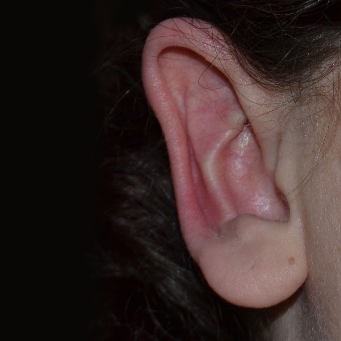
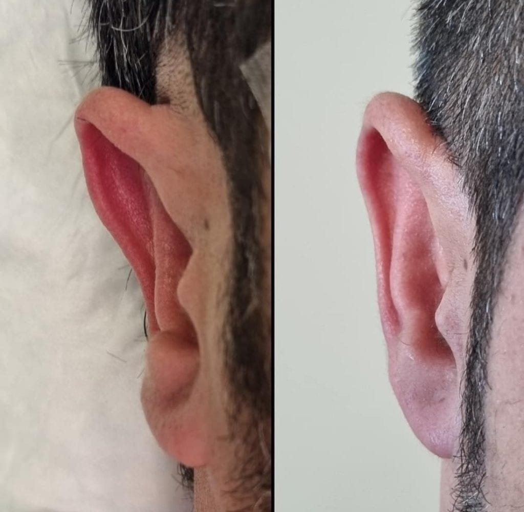
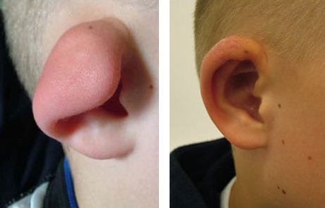
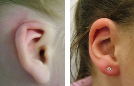
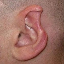
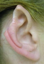
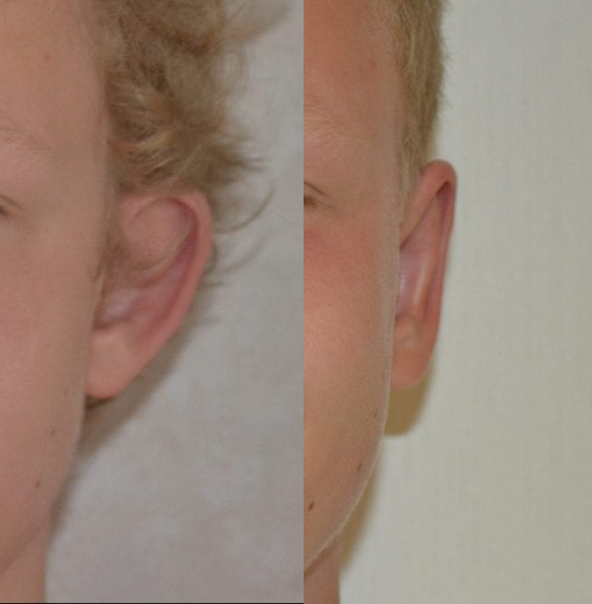
Ear Lobe reduction surgery is an operation to reduce the size of the ear lobe. The ear lobe is a focal point in the appearance of the external ear and is very important in the aesthetics of the ear. Large ear lobes are developmental and hereditary in many cases. Aging also contributes to earlobe enlargement as earlobes become more pendulous with increasing age. Another factor is excessive wearing of heavy earrings which can stretch the earlobes increasing their apparent size. The aim of earlobe reduction is to create a new balance in the size of the ear which in turn will improve the contour of the face.

Am I suitable for Ear Lobe Reduction Surgery?

Earlobe reduction can be carried out on anyone with excessively large ear lobes which are out of balance to create a more youthful earlobe shape. It is a fairly minor procedure and can performed in isolation or as part of a total ear reduction. There are a number of methods to reduce the size of earlobes and choosing the most suitable method will depend on the shape and size of the earlobe. Earlobe reduction is not recommended for patients who have a history of bad scarring or keloids. Also, patients on blood thinning medication may need to stop for 48 hours under the directive of their specialist.

How is Ear Lobe Reduction Surgery performed?

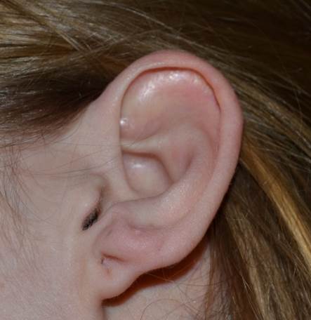
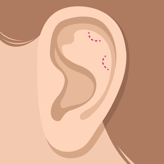
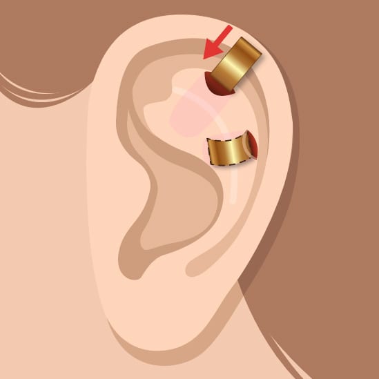
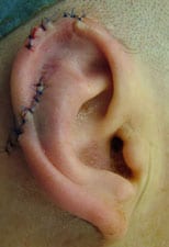
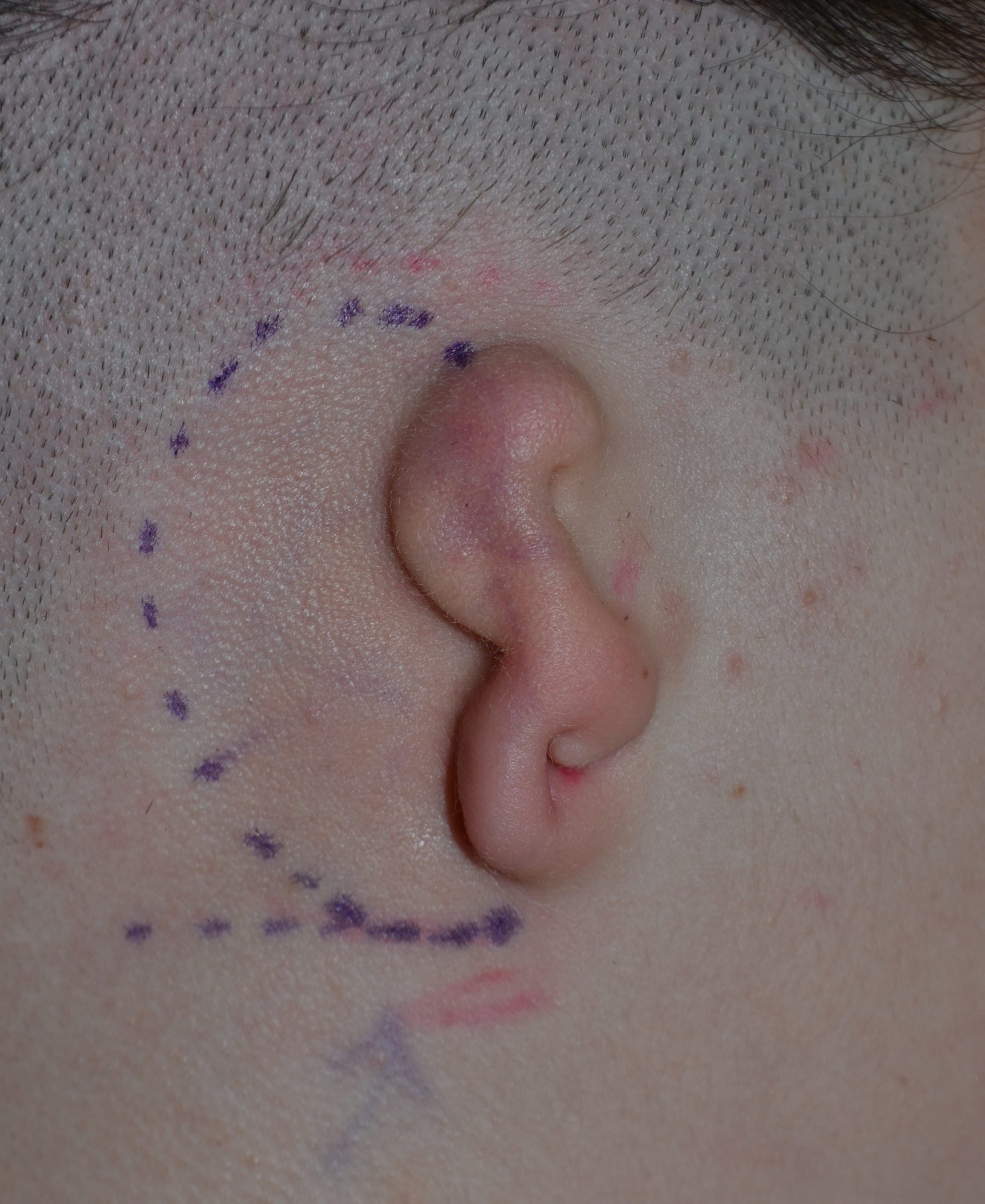
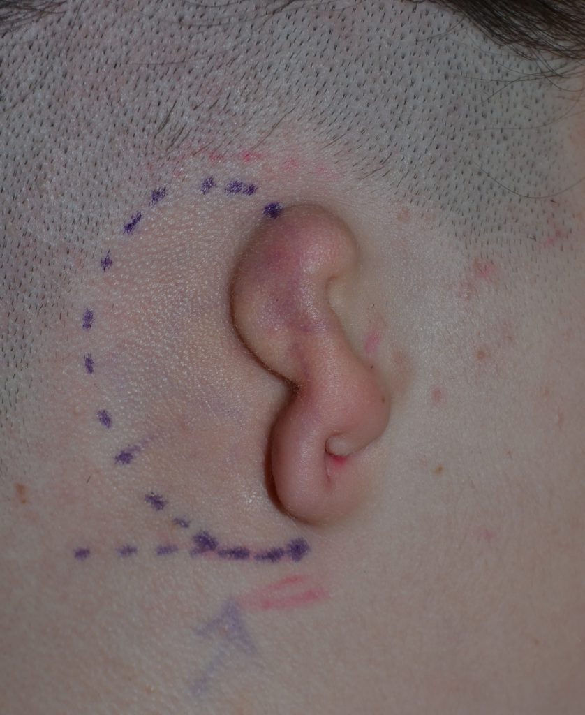
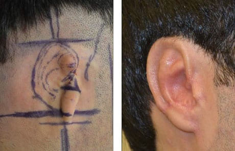
Earlobe reduction is performed under local anaesthetic as a day case procedure. Precise marking of the area to be removed prior to surgery is essential. There are two main methods to reduce the size of the earlobes. The first is resection of the peripheral margin of the earlobe. This leaves a hidden scar on the underside of the earlobe which becomes almost invisible with time. The second method is a wedge reduction usually at the junction of the ear and the face. Both techniques produce aesthetically pleasing results. Selecting the appropriate technique will depend on the shape of the earlobe and the desired outcome. This is assessed at the time of the consultation. Once the excess tissue is removed the wound is closed with dissolvable stitches and a simple dressing applied.

Is Ear Lobe Reduction Safe?

Earlobe reduction is very safe. The most important step is the marking before surgery as the reduction is permeant and therefore precise measurements are needed. Infection risk is minimal and minor bleeding after surgery is of no consequence. The scars resulting from both techniques heal extremely well and will be almost invisible after a few months.

What are the risks in Ear Lobe Reduction?

As in any surgical procedure, complications can occur. In the case of earlobe reduction patients need to be warned of hypertrophic scar, red thickened scar. These occurs in around 2% and can be improved with simple scar management steps. There can be some minor discomfort and itching during the healing period. Minor asymmetry can occur in any earlobe reduction procedure.

What happens after Ear Lobe Reduction?

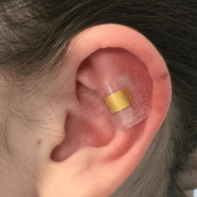
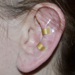
Following surgery, the stitches are covered with very simple adhesive dressing and need to be kept dry for one week. Simple analgesia may be needed for the first few days and patients are asked to avoid putting direct pressure on the ears. Normal activity can be resumed the next day. It is advisable to avoid heavy strenuous activity in the first week as this can increase the swelling. The stitches used are dissolvable and are very fine. The visible pat of the stitch will fall out on the third week. The scar will be red at this stage and will mature and become pale over a period of 3-6 months.

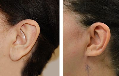
Will I be able to see the results immediately after ear lobe reduction?

You can see the result of the surgery immediately upon completion of the procedure before the dressing is applied. Once the dressing is applied the change in shape and size will still be evident. The dressing is removed at 1 week and there would be very fine stitches still visible at this stage. These will fall out on the third week and at this stage the result of the surgery will clearly visible with very minor swelling which will reduce over the next 4 weeks.


Contact Walid Sabbagh

To arrange an appointment or for any other enquiries, please call or use the form.

Mob: +44 (0)7761 792 835
Tel: +44 (0)203 0020124

  • This field is for validation purposes and should be left unchanged.

Ear Fold

Ear Fold

Ear Fold

The Earfold is a new technique for correcting prominent ears. It is an implant that is introduced through a small incision on the front of the ear. It is placed on the antihelix part of the ear, grips the ear cartilage and immediately adopts a pre-set shape. The Earfold consists of a thin strip of metal (Nitinol) plated with 24-carat gold. Its insertion is a quick procedure performed as an outpatient requiring minimal dressing.

Effective

The Earfold is very effective tool and simple to apply. It is, however, not suitable for all types of ear prominence as it only has one mode of action which is to fold the antihelix of the ear at a preset strength. It can only be applied in cases where folding the antihelix at a specific point gives the desired look. Also it has no effect on the conchal cartilage (the bowl of the ear) which in some cases also contributes to the prominence.

Trial

You will be advised by Mr. Sabbagh of your suitability for the ear fold after full assessment and trial with an implant that is placed above the skin to simulate its effect (see image above).

How it Works

One or two small incisions are made.
The implants are inserted flat, they then spring back to their natural curved shape, which bends the ear back against the head.

What is an Earfold?

Earfold is an implant created to correct prominent ears by reshaping the anti-helix. It is introduced through a small incision under the skin of the ear and grips the ear cartilage and immediately adopts a pre-set shape. The earfold is made from Nitinol alloy (titanium and nickel). The implant consists of a thin strip of metal plated with 24 carat gold to limit its visibility through the skin. No dressings are needed following correction and return to most activities is possible after one week.

How are Earfolds inserted?

Insertion of Earfold implant is a quick and easy procedure performed under local anesthetic. A small incision (5mm) is made within the rim of the ear and the implant (which is pre-mounted on an introducer) is released to correct the prominence. One or two dissolvable sutures are needed to close the incision.

Can Earfolds be used on protruding ears?

The Earfold implant is designed to re-shape the antihelix. If the prominence of the ear is primarily as a result of conchal bowl prominence then the earfold will not correct this and a different surgical procedure will be required.

How long is the recovery period?

No bandage is necessary following Earfold insertion. There will be minor bruising and swelling following its insertion which will subside over a period of 2 weeks.

Is there any risk with an Earfold?

The implant itself has no associated health risks. The implant can be felt under the skin and is almost invisible.

There is a very small risk of infection and in the majority this settles with antibiotics. In rare situations (<1%) the implant may come through the skin. If this happens then the implant has to be removed and the skin heals without any problems. The ear prominence may re-occur after this, especially if this happens within 6 months of insertion, and thus a further procedure may be required.

View before/after photos

Contact Walid Sabbagh

To arrange an appointment or for any other enquiries, please call or use the form.

Mob: +44 (0)7761 792 835
Tel: +44 (0)203 0020124

  • This field is for validation purposes and should be left unchanged.

Ear Reduction

Macrotia Surgery / Ear Reduction

What is Macrotia?

Macrotia is excessive enlargement of the external ear. This refers to the vertical height and width of the ear. The average vertical height for a female ear is 5.9cm and for a male ear 6.2 cm. Usually when the vertical height exceeds 6.5 cm the ear can appear enlarged and out of proportion to the head. Age, sex, height and head size all play a part in how large the ear may appear. Excessively large ears can be a significant cause of concern for many people as they can attract excessive attention and unbalance the harmony of the face.

In the majority of cases the enlargement is in the upper third of the ear as a result of a wide scapha. The scapha is the flattened area between the helical rim and the antihelix. This is the part of the ear that is most commonly reduced in ear reduction surgery. This gives more balance to the ear and better harmony to the face.

Macrotia Causes and Symptoms

There are several pathological conditions that are associated with increased ear size but these are very rare. Age is also a contributing factor with ear circumference increasing marginally as people get older as well as elongation in the earlobes. However, nearly all cases presenting for aesthetic reasons have no specific cause with developmental and genetic factors being primarily responsible. There are no specific symptoms attributed to an increase in ear size. Patients may complain ears are sensitive and uncomfortable wearing certain type of helmets. The main problem is that Macrotia can have a negative effect on confidence and social interaction.

Am I suitable for Macrotia Reduction Surgery?

Nearly all macrotia ears can be reduced in size. Some limiting factors include previous surgery and scarring that can compromise the blood supply to the ear. In such cases ear reduction surgery can still be undertaken by a surgeon who is experienced in ear surgery. Ear reduction in smokers is associated with increased risks in wound healing and it is recommended to stop smoking for 6 weeks before ear reduction surgery.

How is Macrotia Surgery performed?

Different parts of the ear can be enlarged disproportionally and surgical treatment is individually tailored.  Macrotia surgery is usually performed by removing a crescent of skin and cartilage from the scapha on the front of the ear. Skin on the back of the ear is not removed. The width of the crescent will dictate how much the ear is reduced. The outer arc of the crescent is then advanced towards the inner arc to close the gap. This usually necessitates removal of a small segment of the helical rim. The resulting scar is just within the helical rim a small horizontal scar on the rim itself. These scars heal extremely well that they are almost invisible after a few months.

The surgery can be performed under local anaesthetic and all the stitches are dissolvable. A bandage is worn overnight but can be removed after 24 hours.

Is Ear Reduction Safe?

Ear reduction surgery is a very safe operation when performed by an ear specialist as it requires experience and appreciation in how the ear alters its shape during the reduction process. When the operation is performed with focus on precision, meticulous technique and appreciation of ear skin blood supply complications are negligible. In addition, by performing the procedure under local anaesthetic risks from a general anaesthetic, although extremely rare, are also eliminated.   

Ear Reduction Surgery Risks

Complications that can occur following ear reduction are rare and minor. There is a minor degree of swelling which usually resolves by the second week. In the first 24 hours minor bleeding from the wound may occur which is of no consequence. Infection risk is very small (1%) and responds well to antibiotics. Scars can be slightly red in first 3 months but always settle down.

There is an increase risk complication in smokers and it is recommended to stop smoking for 6 weeks before surgery as nicotine constricts the blood vessels decreasing the blood supply.

Before and After

The image to the left shows an example of the before and after of the Ear Reduction surgery.

More images are available in the click at the bottom of the page.

What happens after ear reduction surgery?

At the completion of the operation antibiotic ointment is applied to the wound and soft cotton wool placed over the stitches. A bandage is then applied which will hold the dressing in place over for the first night. The bandage is removed the next day and a very soft head band is provided to cover and protect the ears. There is minor discomfort following the operation which is well controlled with paracetamol / ibuprofen. Most gentle daily activities can be resumed the next day. It is best to delay vigorous physical activities for 2-3 weeks until the wounds have completely healed. Following wound healing the ears may remain slightly tender to touch for a few months as the scar remodels. After 6 months the scar is almost invisible.

Will Surgery affect my hearing?

Ear reduction surgery does not interfere with the hearing mechanism of the ear.

When will I see the results of ear reduction surgery?

You will be able to see the results of ear reduction surgery immediately in the operating theatre at the completion of the operation before a bandage is applied. The bandage can be removed the next day and the result of the reduction will visible but at this stage there will be some cotton wool over the stitches in the scapha and it is important to appreciate that at this stage there is some swelling. The swelling will reduce over 2 weeks and the stitches will fall out on the third week.  

Contact Walid Sabbagh

To arrange an appointment or for any other enquiries, please call or use the form.

Mob: +44 (0)7761 792 835
Tel: +44 (0)203 0020124

  • This field is for validation purposes and should be left unchanged.

Revision Otoplasty

Revision Otoplasty

Why would you need revision otoplasty surgery?

There are a number of reasons why there could be an unsatisfactory outcome following Otoplasty requiring revision surgery. These include:

  • Insufficient correction / recurrence of ear prominence
  • Poor cosmetic outcome and / or asymmetry
  • Inappropriate otoplasty: this occurs when the patient undergoes an ear pinning procedure in a macrotia (large ear). The result is usually unsatisfactory as in these cases an ear reduction procedure was needed.
  • Overcorrection of a protruding ear. This usually happens in the mid portion of the ear resulting in what is known as a telephone ear deformity. In the majority of these case a strut of rib cartilage is needed behind the ear to undo the over-correction
  • Infection and cartilage loss resulting a significant deformity. These are rare and are likely to require rib cartilage reconstruction

What is Revision Otoplasty?

Revision Otoplasty is an operation to the ears applied to remedy the problems outlined above following unsatisfactory Otoplasty surgery. These problems cause significant patient distress and in most cases the patients become more anxious about their ears than they were prior to the original Otoplasty procedure. Revision Otoplasty is not one type of surgery but a number of techniques designed to improve the ears appearance. It does not aim to reverse the original Otoplasty as in nearly all cases this is not possible. However, it will address the specific problems and set objectives that can improve the ear appearance.

Am I suitable for Revision Otoplasty?

In nearly all patients who are unsatisfied with their otoplasty outcome a revision procedure is an option. The type of revision otoplasty that may be needed will require a consultation to assess the ear and a detailed discussion of what surgery can achieve. A simple reversal of an unsatisfactory otoplasty is rarely possible, however, in the majority of cases a revisional procedure can improve the appearance of the ear and thus the confidence of the patient.

Is Revision Otoplasty Safe?

Revision otoplasty is very safe and in cases where rib cartilage is not needed then nearly all can be performed under local anaesthetic. Performing surgery on an already operated on ear requires a meticulous approach in view of the scar tissue that is already present. This, however, does not mean there is an increased risk of post-surgical complications. With care and precision healing is relatively straight forward with minimal complications. Minor complications that can occur include swelling, bruising and numbness which all would settle over a period of weeks.

When rib cartilage is required for the revision then a general anaesthetic is needed. This can still be done as a day case procedure. The harvest of rib cartilage is extremely safe and the patient is mobile from the next day needing some pain killers for a period of 5-7 days.

Revision Otoplasty Risks

Revision otoplasty encompasses a number of different procedures. In revision otoplasty there is scar tissue from previous surgery. Operating in an area of scar tissue slightly increases swelling post-surgery.  Scar tissue is also very vascular and this can cause minor bleeding in first 24 hours after surgery but this is rarely of any consequence. Infection risk is very small (1%) and antibiotics are given in revision surgery. It is always more difficult in revision surgery to attain the desired cosmetic outcome and this would be communicated in detail in the consultation. If rib is required there would some discomfort at the site of the rib harvest which usually settles by two weeks.

What happens after Revision Otoplasty?

At the completion of the operation a bandage is applied to protect the ears for the first night. This can be removed the following day. Pain killers (Paracetamol and Ibuprofen) are usually needed for the first few days. It is advisable to keep the wound dry and showering can commence after day 5. Most normal daily activities can resume after a few days. Heavy activities / sport can re-start after 2 weeks when most of the swelling has subsided. Following wound healing the ears will remain slightly tender to touch for a few months as the scar remodels.

When will I see the results after Revision Otoplasty?

You will be able to see the results of revision otoplasty surgery on the same day the procedure is performed. A bandage will be applied in theatre and is changed on the ward prior to discharge. The result of the surgery will visible from the next day when this bandage is removed. It is important to appreciate that at this stage there will be some swelling. This will reduce over 2 weeks and the stitches will fall out on the third week. 

View before/after photos

Contact Walid Sabbagh

To arrange an appointment or for any other enquiries, please call or use the form.

Mob: +44 (0)7761 792 835
Tel: +44 (0)203 0020124

  • This field is for validation purposes and should be left unchanged.

Ear Keloids / Lumps

Ear Keloids / Lumps

Unsightly and uncomfortable lumps are common on the ear which include Keloids and Cysts. Other lesions that can occur on the ear are benign moles, skin tags and skin cancers as well as skin irritation secondary to underlying inflamed cartilage.

Options

Keloids are excessive scar production by the body and most often occur at the sight of piercing. There are a number of options available to treat keloids involving injections and surgery.

Other swellings

Other problems that may require removal from the ear include cysts, chondrodermatatis (inflammation in cartilage) and various types of skin cancers. Mr Sabbagh will remove all the swellings around the ear with focus on maintain the normal shape of the ear and optimum scarring.


View before/after photos

Contact Walid Sabbagh

To arrange an appointment or for any other enquiries, please call or use the form.

Mob: +44 (0)7761 792 835
Tel: +44 (0)203 0020124

  • This field is for validation purposes and should be left unchanged.

Other Ear Abnormalities

Other Ear Abnormalities

Constricted ears

The main characteristic of this abnormality is a fold in the upper portion of the ear. Other terms used to describe this deformity are constricted ear and lop ear. Other features that are often associated with cup ears include shortening and prominence of the ear. The type of surgery performed for cup ears depends on the severity of the abnormality.

For mild cup ears with a small fold, the extra cartilage and skin can simply be removed. In moderate folds, the folded cartilage is removed and re-inserted in a reverse direction. Permanent sutures can also be placed at the back of the ear to correct the abnormality. In severe cup ear deformity there is a shortage of skin and cartilage and rib cartilage will need to be inserted to re-define the rim.

Cryptotia

Cryptotia refers to an ear that appears to have its upper portion buried underneath the side of the head.

Cryptotia is also known as buried ear or hidden ear.

Cryptotia is often treated through surgery which releases the ear from its buried position, reshaping the cartilage and using local tissue to resurface the released cartilage.


View before/after photos

Contact Walid Sabbagh

To arrange an appointment or for any other enquiries, please call or use the form.

Mob: +44 (0)7761 792 835
Tel: +44 (0)203 0020124

  • This field is for validation purposes and should be left unchanged.

Reconstruction after Trauma

Reconstruction after Trauma

Partial or complete traumatic ear loss can occur as a result of various injuries. The most common cause in the UK is related to physical assault through human bites. Other causes include infection following piercing, which can destroy some of the ear cartilage, accidents, cancer surgery and burns.

Reconstruction

In nearly all cases of ear loss the missing part of the ear can be reconstructed using sculpted rib cartilage. A three dimensional framework of the missing segment is carved and positioned under the adjacent skin to reconstruct the ear. This is a complex operation which Mr Sabbagh has performed on hundreds of cases with excellent outcome.

Procedure

The reconstruction of the ear is performed in two stages. In the first stage the shape of the ear is recreated. This results in a normal looking ear, however, because of the shortage of skin the reconstructed ear will be attached to the side of the head and require separation in the second stage. This is performed six months after the first stage. The ear is elevated and a skin graft is applied to cover the back of the new ear.

Example of traumatic ear loss
A three dimensional framework
Post operation result


Contact Walid Sabbagh

To arrange an appointment or for any other enquiries, please call or use the form.

Mob: +44 (0)7761 792 835
Tel: +44 (0)203 0020124

  • This field is for validation purposes and should be left unchanged.

Microtia

Microtia

Microtia is a rare congenital abnormality of the ear. The term Microtia means ‘small ear’ and occurs one in every 6000 births.

Microtia is categorised according to its appearance:

  • Lobular Microtia: The most common, only the lobe of the ear present
  • Conchal Microtia: The lower third of the ear has a normal appearance
  • Anotia: The whole ear absent
  • Atypical types: Cases that do not come under above categories

Reconstruction

The reconstruction process is in two stages, about 6-12 months apart. The first operation involves re-orientating the available tissue and thinning the skin to mimic the ear skin. Rib cartilage (using ribs 6, 7, 8 and 9) is then harvested and carved to produce a very detailed framework. The framework is inserted in the pocket and gentle suction is applied to enable adherence of skin. This operation takes 4-6 hours and expected length of stay is 3-5 days. At the completion of this stage the whole ear will be visibly reconstructed but will be adherent to the side of the head.

The second stage involves releasing the ear from the head and inserting a piece of cartilage behind it to maintain projection. The cartilage is covered with vascularised tissue and a skin graft. This takes 3-4 hours and expected hospital stay is 2-3 days.

Mr Sabbagh is one of the world experts in ear reconstruction for Microtia and has been performing this complex surgery for over 10 year with experience in several hundred cases. The timing of surgery in children is usually around 9-10 years old, when the rib cartilage is large enough, but can also be performed in adults. Excellent results can be achieved with ears that have sensation and durable throughout the patients’ life.

Contact Walid Sabbagh

To arrange an appointment or for any other enquiries, please call or use the form.

Mob: +44 (0)7761 792 835
Tel: +44 (0)203 0020124

  • This field is for validation purposes and should be left unchanged.

Ear Pinning / Ear Stitch

Ear Pinning

Ear Pinning

Ear pinning is the most common reason to undergo corrective ear surgery. Correction of prominent ears creates better harmony to the face and can result in a considerable improvement in the psychological wellbeing patients. The aim of surgical ear pinning is to position the ears closely aligned with the head but in a natural position so they blend with the face rather than being a significant focus in facial appearance. This improves the balance of the face and the overall appearance of other facial features. As well as optimising facial aesthetics ear pinning considerably improves self-esteem and confidence.

Stitch technique

Mr Sabbagh uses the stitch technique, also known as ear stitch, to correct prominent ears. The stitches enable accurate movement of different parts of the ear and thus the upper, middle and lower third can be pinned back to the degree the patient desires. With this technique the ear position is completely controlled thus achieving a natural look with a naturally curved antihelix and a visible helical rim on frontal profile. This technique is very effective and predictable. The surgery is performed under local anaesthetic from behind the ear and the resulting scar is almost invisible.

Preparing for the procedure / what to expect before during and after

The ear stitch technique requires minimal preparation. As it is performed under local anaesthetic no fasting is needed before the surgery. Patients are advised to wash their hair the night before. On admission (one hour prior to the procedure) the nursing staff will carry out routine assessment and Mr Sabbagh will attend to confirm all the points that were discussed at consultation. The environment in the operating theatre is very relaxed and the nursing staff will always make the patient feel very comfortable. Local anaesthetic is injected behind the ear to numb the ear and surgical drapes are placed to create a sterile environment. The surgery takes around 30 minutes per ear and on completion the patient is able to inspect the ears with a hand-held mirror. A bandage is applied at completion, as a dressing, which is removed the next day. The patient is discharged following a review one hour after surgery. Pain medication will be provided.

How is the operation performed?

Surgery is performed from behind the ear. An incision is made in the skin to gain access to the ear cartilage. The fold of the cartilage visible on the front of the ear (antihelix) is recreated by inserting non-dissolving stitches in the cartilage. As the stitches are tied very accurate manipulation of ear position is possible. This allows a very natural result to be achieved. In some ears the conchal bowl (the deep concavity of the ear) is excessively prominent. This is repositioned with sutures or, if very stiff, a small crescent of the conchal cartilage is removed. The skin is then closed behind the ear with dissolvable sutures.

From what age can the surgery be performed?

Correction of prominent ears can be performed at any age after 5 years. Although it is possible to perform the surgery before the age of 5 it is not advisable as the operation requires understanding and co-operation from the patient during and after the surgery. It can be performed under local anaesthetic at any age, however, for children under the age of 10 general anaesthesia is usually preferred.

Is the operation painful?

There will be some localised pain around the ears and this is alleviated by basic painkillers such as paracetamol and brufen. This significantly improves after 24 hours and fully resolves within a week. It is recommended to take regular pain killers for the first few days; after then they can be taken as required. Most patients are able to resume their daily activities around two days post-surgery. Although not painful, the ears can remain slightly tender to touch for 3 months following the surgery.

Anaethesia

The ear stitch is a minimalist technique and can easily be performed under local anaesthetic. This has many advantages as it eliminates any risks from a general anaesthetic, has quicker recovery and allows the patient to view the result at completion. The local anaesthetic used is the same as by the dentist and ears are numbed by a quick injection behind the ear. The injection is very straightforward to administer and causes minimal discomfort. For small children general anaesthetic is usually preferred as co-operation is needed during the operation. However, if the child is of a mature age local anaesthetic is also an option. The effect of the local anaesthetic lasts for several hours so there is no concern that its effect may wear off during the procedure.

Are there any risks?

This is a procedure with a very high level of satisfaction, very safe with minimal risks. Complications are very rare and minor. There may be some bleeding from the wound which is of no consequence. There will be some swelling which subsides by the second week. Infection rate is very low, less than 1%. In the majority scars heal very well and are almost invisible. In a small group of the population thicker (hypertrophic) scars may form which may need further treatment. There is a very small risk (less than 5%) of partial recurrence of the ear prominence. This is most likely to occur in the first six months and can be easily corrected if necessary.

Recovery

Recovery is quick with no bruising on the front of the ear and return to work is possible after a few days and children to school after one week. A bandage is not necessary but can be worn for comfort for first 24 hours. Analgesia is likely to be needed for the first week. Most activities can be resumed after 48 hours. There will be some swelling and most of this will disappear by the second week. Most physical activities/sport can be resumed after the second week. For contact sports such as rugby or martial arts it is advisable to wait 4 weeks and to carry on protecting the ear with an elastic headband for 3 months. Swimming can be resumed after the third week.

Will there be scars?

There will be a surgical scar behind the ear which will go through several stages of healing. The dissolvable stitches in the skin fall out on the third week. The scar will appear as a faint line for around 6 weeks and will eventually become invisible. In very small percentage the body can form excessive scarring / keloids. These can be managed accordingly.

Will I need to wear a bandage?

A head bandage covering the ears and forehead only will be placed at surgery. The aim of the bandage is to make healing more comfortable and minimize minor bleeding that may occur. The bandage can be removed the next day as dictated by patient’s comfort. The patient will be advised to wear a headband at night and during sport activities for 6 weeks. The bandage has no functional role to keep the ears in position. It is worn for comfort and protection of the ear when needed.

Gallery

View before & after photos of Mr Sabbagh’s work.

Contact Walid Sabbagh

To arrange an appointment or for any other enquiries, please call or use the form.

Mob: +44 (0)7761 792 835
Tel: +44 (0)203 0020124

  • This field is for validation purposes and should be left unchanged.